 |
Intelligent Medical Information Computing Laboratory
|
Dalian University of Technology Department of software engineering |
| Lab Home
|
People
|
Research
Projects
|
Publications
|
Join Us
|
Localization and Segmentation of Human Fetal Brain Ultrasound Image
|
||||||||
Motivation
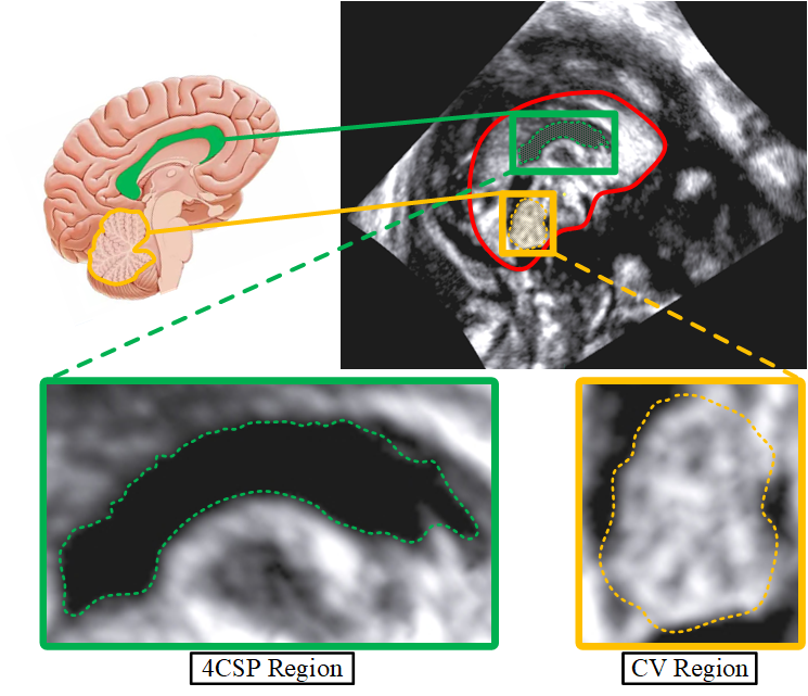
Newborns are born every day all over the world, but unfortunately, some of them are not healthy. With increased awareness of the importance of prenatal care and medical advances, preconception and prenatal examination have become essential for gravidas before giving birth. In the prenatal examination process, it is important to inspect the development of the fetal brain. Among the many aspects of the fetal brain examination, the examinations of the Corpus Callosum (CC) and Cerebellar Vermis (CV) are critical. The 20th to 28th gestational weeks are the best time period for these examinations. During this time, the CC and CV have formed and there is space in the cranial cavity to facilitate ultrasound imaging. Noteworthy, in fact that physicians diagnose the developmental status of CC by observing the Complex of Corpus Callosum and Cavum Septum Pellucidum (4CSP) in fetal brain mid-sagittal ultrasound images (FBMUIs). Therefore, the main research subjects are 4CSP and CV in FBMUIs.
|
||||||||
The 4CSP and CV region in FBMUI |
||||||||
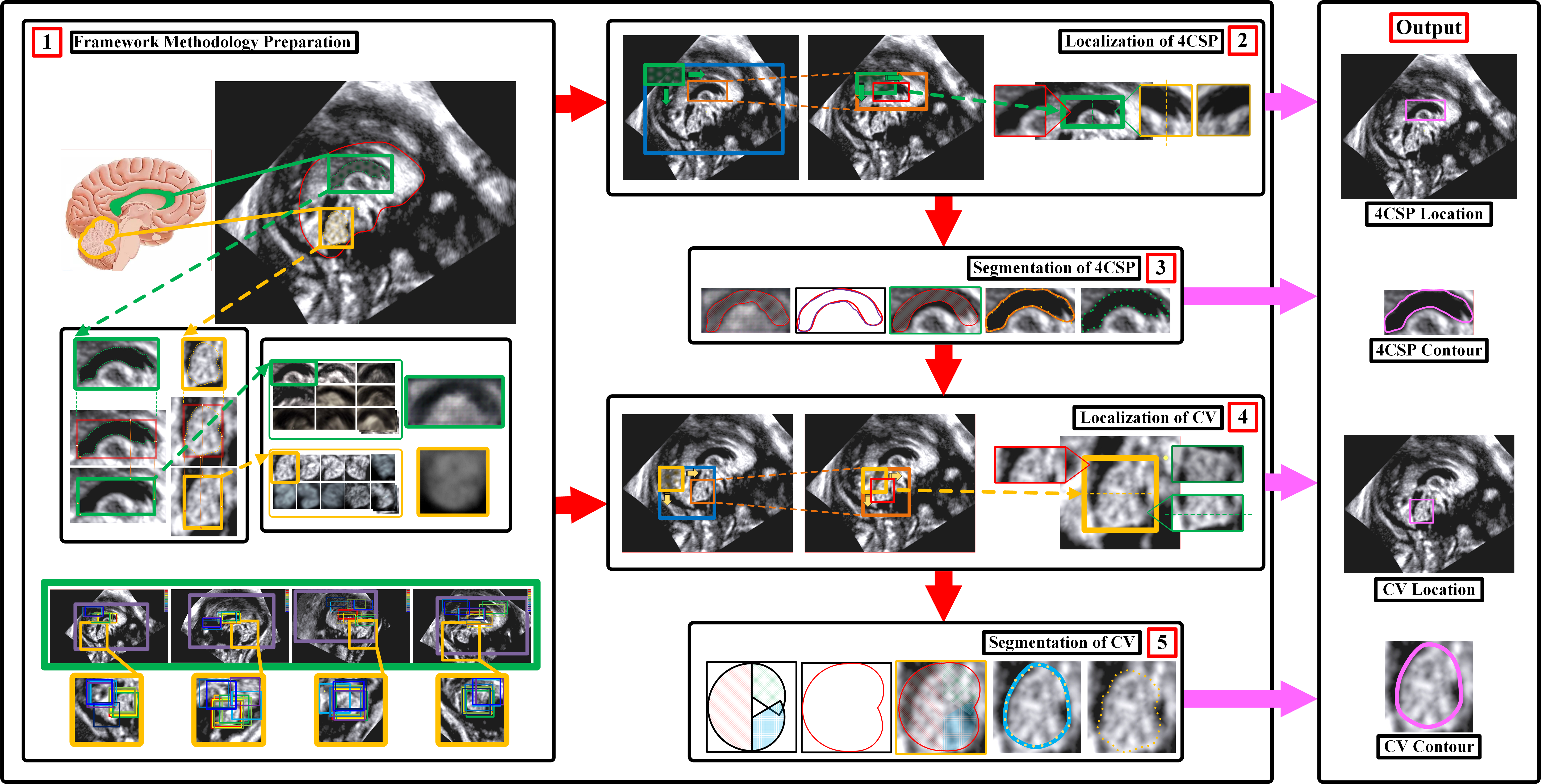
Flowchart of localization and segmentation of 4CSP and CV in FBMUIs
We present an integrated framework for automatic localization and segmentation of the Complex of Corpus Callosum and Cavum Septum Pellucidum (4CSP) and Cerebellar Vermis (CV) in ultrasound images of the fetal brain based on a priori anatomical knowledge and computer vision methods. The framework aims to obtain location and contour information of CC and CV in fetal brain mid-sagittal ultrasound images (FBMUIs) to assist physicians in prenatal screening examination. There has been no study specially addressing this issue and our study has important clinical implications for early diagnosis of human embryos. |
||||||||
Flowchart of localization and segmentation of 4CSP and CV in FBMUIs. The green and yellow dashed lines respectively reflect the correspondence between the steps in the localization and segmentation of 4CSP and CV (dashed lines connect images or rectangles, where the front and back indicate the same image or a derived relationship). The overall framework is a string structure, and the result of the previous step is the input of the next step, including five steps: Generation of the Average Templates; Localization of 4CSP; Segmentation of 4CSP; Localization of CV; Segmentation of CV.
|
||||||||
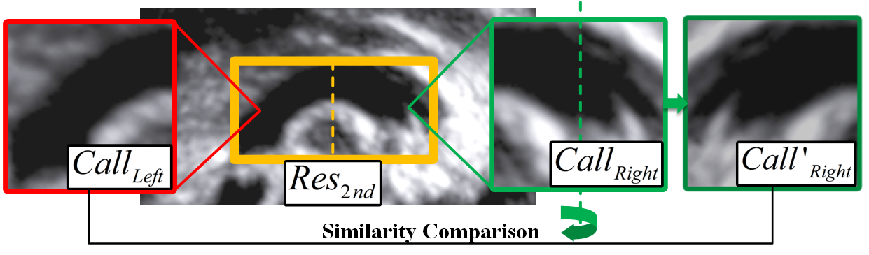
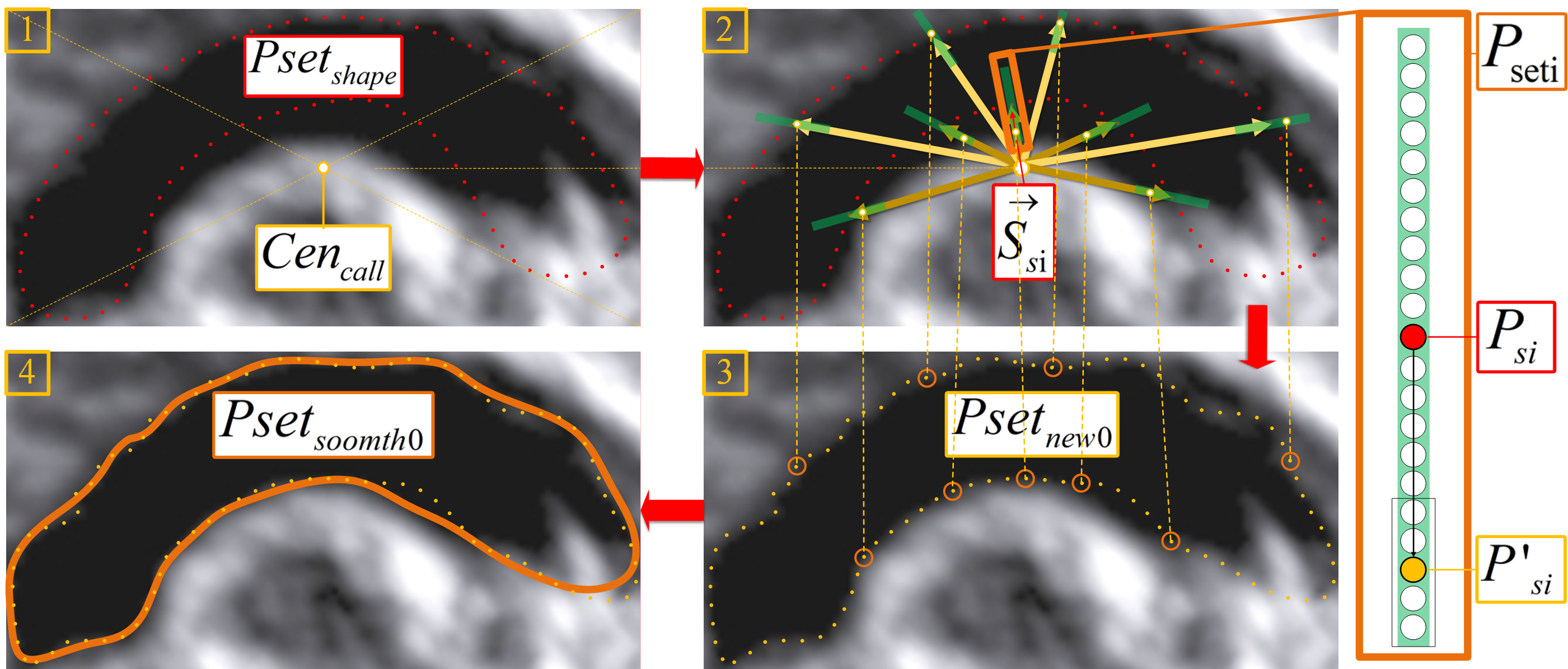
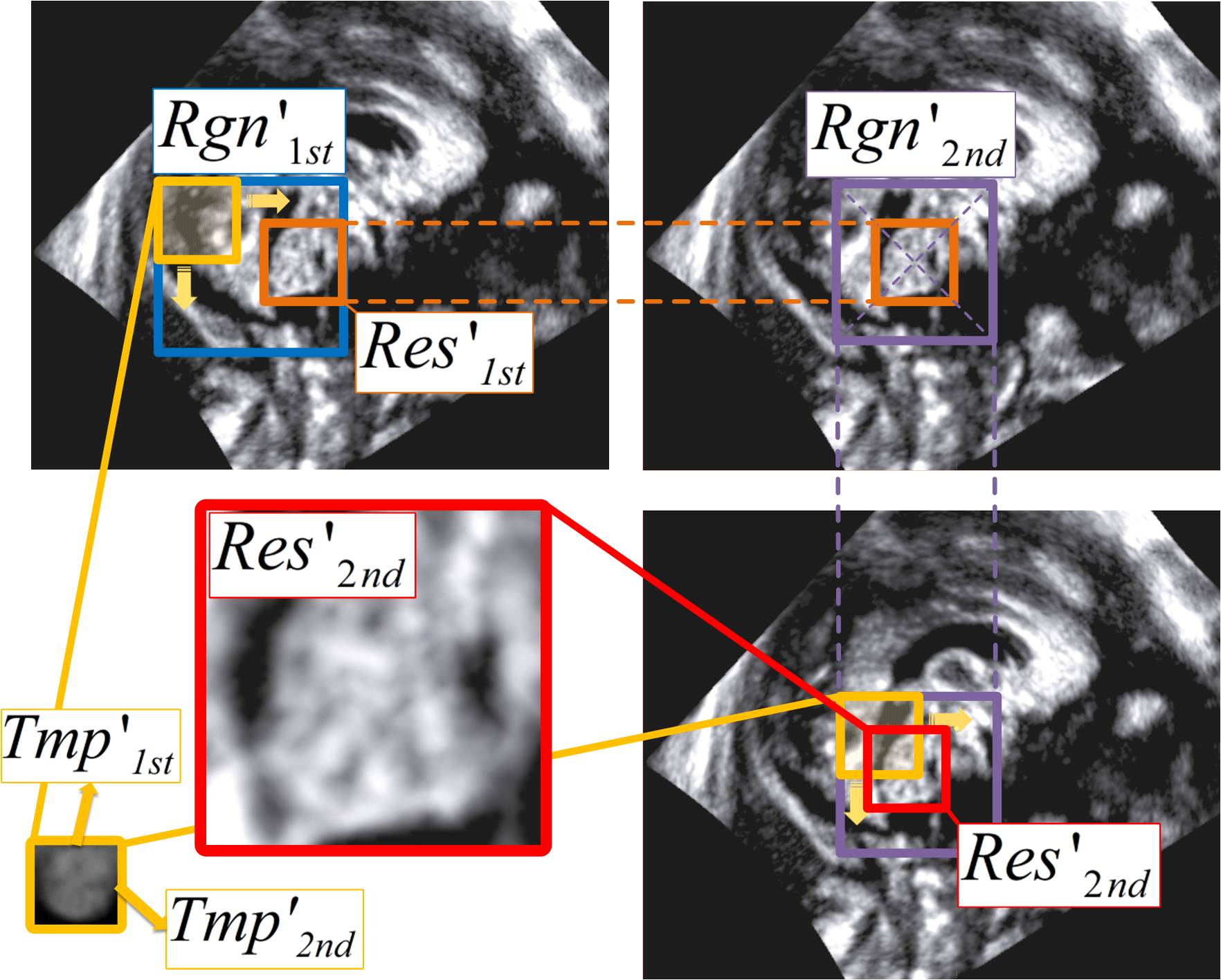
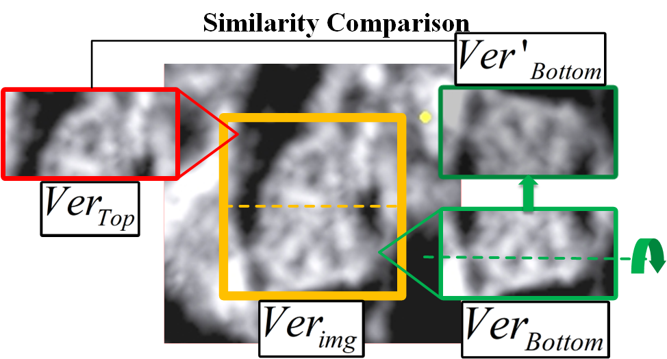
Localization and Segmentation of the 4CSP Region
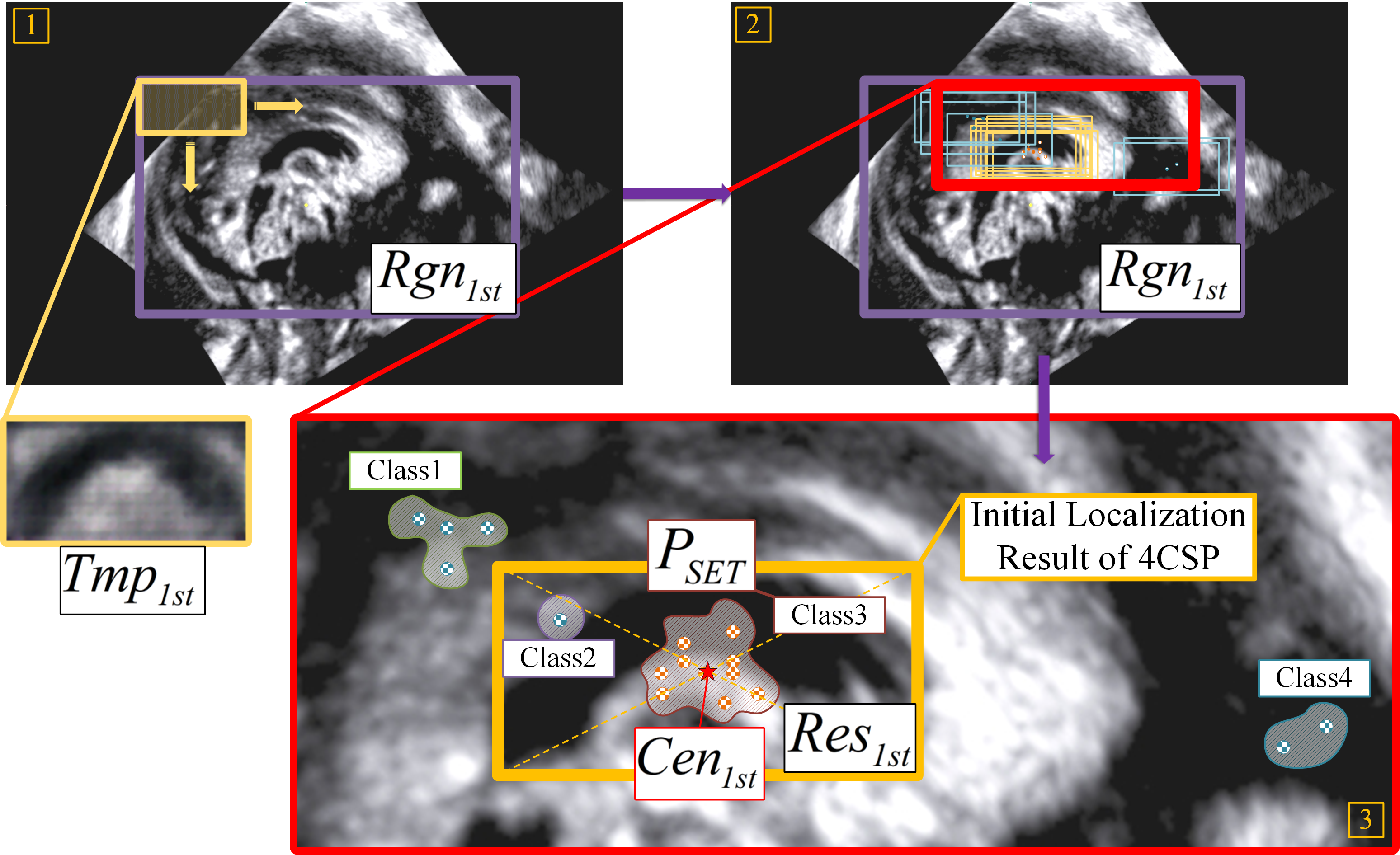
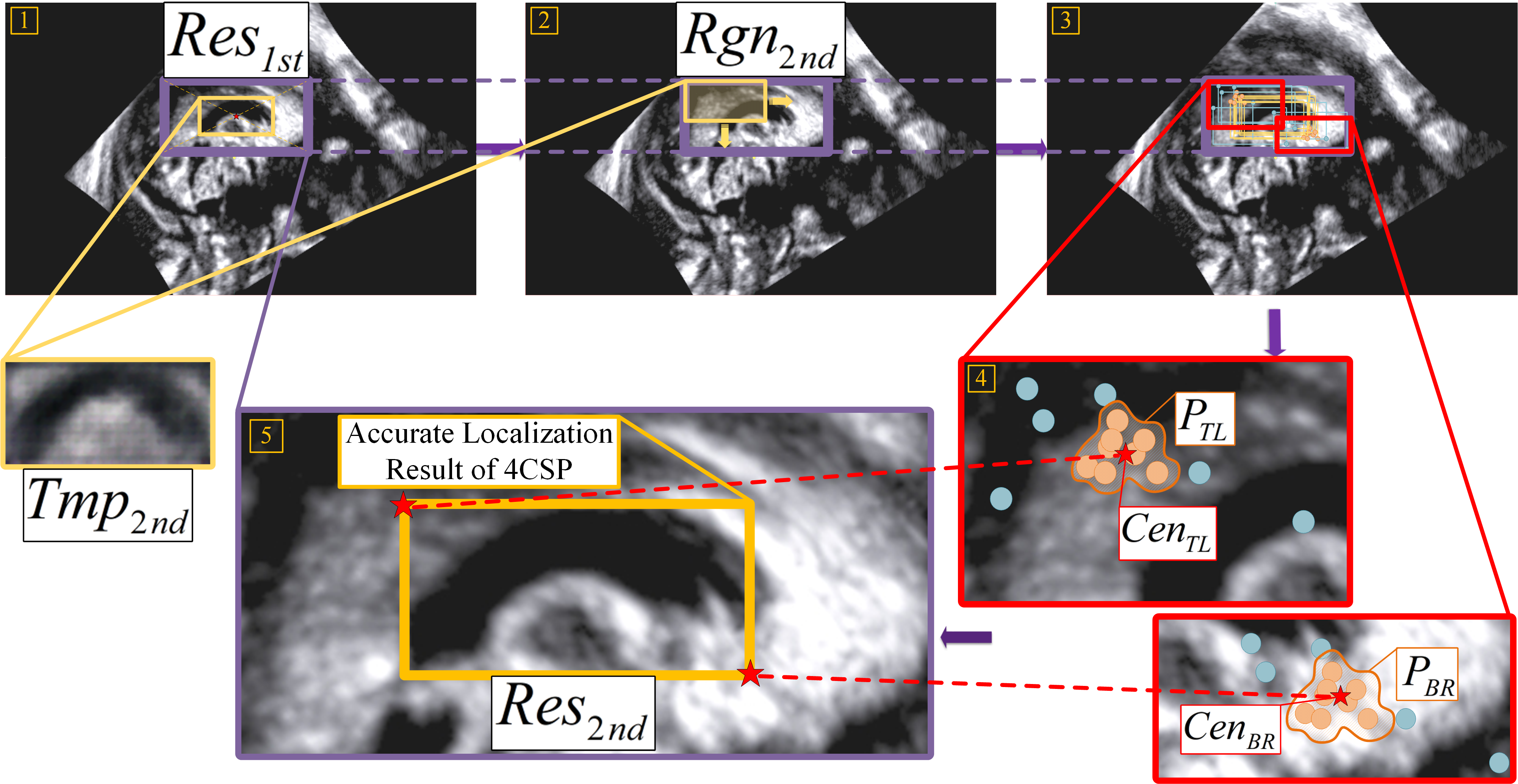
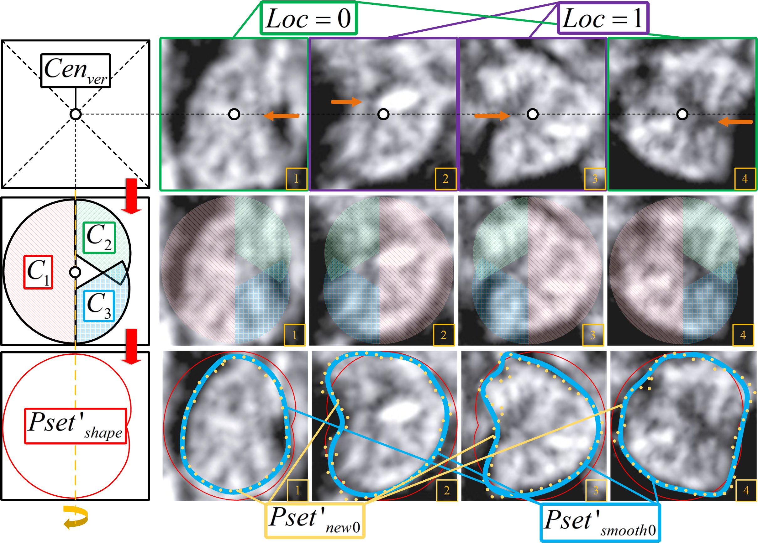
The localization process of 4CSP adopts the sequential structure of "Initial Localization-Accurate Localization-Result Detection", and completes the segmentation of 4CSP through the structure of "Contour Fitting-Contour Iteration".
|
||||||||
Localization and Segmentation of the CV Region
The localization process of CV adopts the same sequential structure as 4CSP: "Initial Localization-Accurate Localization-Result Detection", and completes the segmentation of CV through the structure of "Contour Fitting-Contour Iteration".
All ProcessVideo We have designed and produced software to test the feasibility of the framework. Currently, the method has a good response in clinical experiments in Shengjing Hospital.
|