 |
Intelligent Medical Information Computing Laboratory
|
Dalian University of Technology Department of software engineering |
| Lab Home
|
People
|
Research
Projects
|
Publications
|
Join Us
|
Precise Anchor Location For Cruciate Ligament Surgery
|
||||||||
Motivation
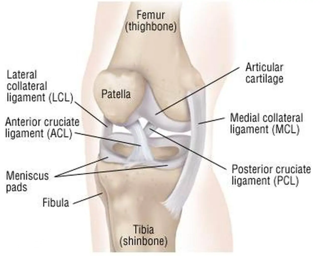
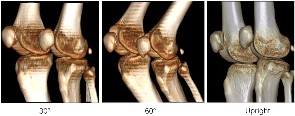
Cruciate ligament connects bone and bone. The role is to strengthen the stability of the joint, so as to avoid injury. When suffering from violence or non-physiological activities, ligaments are stretched beyond their tolerance, resulting in injury.
|
||||||||
|
||||||||
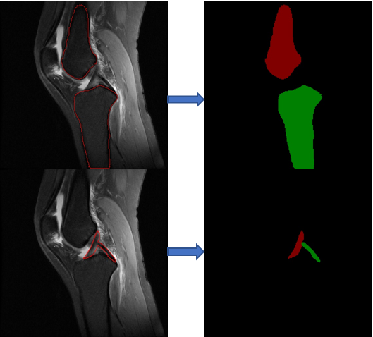
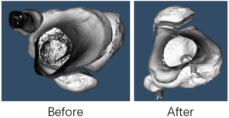
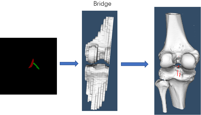
Cruciate ligament injury is very common in daily life. At present, many ligament reconstruction operations are based on the experience of doctors, and errors are inevitable. Extraction and reconstruction of cruciate ligament insertion is very important, which can help doctors to analyze and predict before surgery, and improve the success rate of surgery.
|
||||||||
|